This project is designed to investigate how the release of brain-derived neurotrophic factor (BDNF) from corticostriatal afferents and tropomyosin receptor kinase B (TrkB) responsiveness in striatal medium spiny neurons are regulated by dopaminergic activity from the substantia nigra and by deep brain stimulation (DBS) of the subthalamic nucleus (STN).
The project builds on our expertise with cell culture techniques for enrichment of medium spiny neurons and new mouse models of dystonia, as well as models in which BDNF signaling in these corticostriatal synapses is disturbed. The goal is to understand how plasticity mechanisms at this synapse contribute to motor defects in Parkinson´s disease and dystonia, and how DBS of the STN modulates this synapse and motor function.
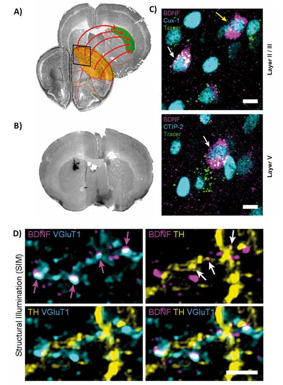
Fig.1 BDNF is expressed in traced cortico-striatal projection neurons: A) Schematic representation of motor cortex projections to the dorsolateral striatum. B) Injection site for fluorescent retrograde tracer beads in dorsolateral striatum. C) IHC of PFA fixed motor cortex brain sections reveal BDNF expression in retrogradely traced cortico-striatal projection neurons in layer II/III (Cux-1) and layer V (CTIP-2). Scale bar: 10µm. D) BDNF-IR in presynaptic terminals in the dorsolateral striatum. BDNF-IR in cortical VGluT1-positive terminals (magenta arrows) versus midbrain TH-positive terminals (white arrows). VGluT1- and TH-positive terminals reside in direct regional proximity but do not overlap. Andreska et al. J. Neurosci. 2020.
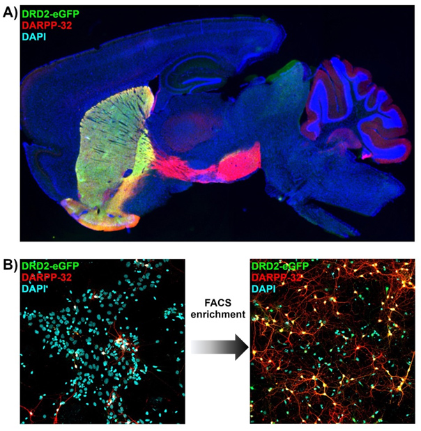
Fig.2 BAC transgenic fluorescent reporter mice for investigation BDNF / TrkB signaling in striatal MSNs : A) IHC of BAC transgenic DRD2-EGFP mouse. Indirect pathway DRD2-MSNs are highlighted in green (eGFP). All MSNs were highlighted with DARPP-32 (red). B) Schematic representation of FACS enrichment of striatal MSNs in vitro. MSN cell cultures derived from P1 DRD2-eGFP mice before (left) and after FACS enrichment of DRD2-eGFP MSNs (right). Images show cultures at DIV7.
Team
Publications
Dopaminergic Input Regulates the Sensitivity of Indirect Pathway Striatal Spiny Neurons to Brain-Derived Neurotrophic Factor
- Maurilyn Ayon Olivas
- Daniel Wolf
- Dr. Thomas Andreska
- Prof. Chi Wang Ip
- Prof. Michael Sendtner
Calnexin controls TrkB cell surface transport and ER-phagy in mouse cerebral cortex development
- Dr. Thomas Andreska
- Daniel Wolf
- Prof. Michael Sendtner
DRD1 signaling modulates TrkB turnover and BDNF sensitivity in direct pathway striatal medium spiny neurons.
Insulin-like growth factor 5 associates with human Aß plaques and promotes cognitive impairment.
- Dr. Thomas Andreska
- PD Dr. Robert Blum
- Prof. Michael Sendtner
Development of a Fully Implantable Stimulator for Deep Brain Stimulation in Mice.
- Prof. Jens Volkmann
- Prof. Michael Sendtner
Regulation of TrkB cell surface expression-a mechanism for modulation of neuronal responsiveness to brain-derived neurotrophic factor
- Dr. Thomas Andreska
- Prof. Michael Sendtner
Induction of BDNF Expression in Layer II/III and Layer V Neurons of the Motor Cortex Is Essential for Motor Learning
- Dr. Thomas Andreska
- PD Dr. Robert Blum
- Prof. Philip Tovote
- Prof. Michael Sendtner


